SUPERCMA, a new class of Cylindrical Mirror Analyzers SUPERCMA is a major improvement of the
classical cylindrical mirror analyzer (CMA). The new SUPERCMA design offers all the advantages of the regular CMA and overcomes all the weaknesses of its predecessor. The working distance is very convenient, ranging from 35 mm to 55 mm depending on the model. Sample access and positioning are much easier, and changes in sample position are more than ten times less critical. The sample distance to the analyzer can be changed by a several millimeters still maintaining all characteristics. The accepted sample area is as large as 3 mm in diameter, eliminating the constraint of using a well focused beam for analysis. This is a major advantage when performing Scanning Auger Microscopy with the analyzer, because the scanned area can be large. In addition, the SUPERCMA offers new features which were until now considered characteristic of only larger, powerful hemispherical systems.
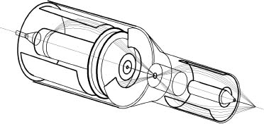
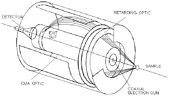
Why SUPERCMA is a superior device: The key to these surprisingly good features is the use of new properties of the cylindrical mirror geometry. In contrast to the standard CMA geometry, where the electron beam enters and exits the cylindrical field passing through slits machined into the inner cylinder, the new super CMA accepts electrons entering between the two cylinders as shown in Figure a). This patented geometry has good focusing properties and a large working distance. Combined with a high precision retarding field optic, the electron energy through the cylindrical mirror field, i.e., the pass energy, is variable and so is the energy resolution.
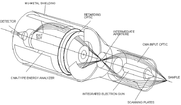
Figure a) Single Pass versus Double Pass The single pass design, as described above, is ideally suited for standard AES investigations in analog and pulse counting modes. A more powerful instrument is obtained combining two CMA fields in series, as shown in Figure b). This double pass geometry uses the first element as an input lens and the second as the energy analyzer. The main advantage of this design is that the sample area as seen by the analyzer is well defined by the input optic. This configuration allows XPS measurements where the sample area is widely exposed to the X-ray beam. In this case, the analyzed area on the sample is well defined and limited to a small circular spot in the millimeter range.
Figure b) Members of the SUPERCMA family
|
|||


